

Learning radiology takes time. There is no shortcut around that. But the resources you choose can make a huge difference in how quickly things start to click. The right mix of websites, books, videos, and case libraries can turn what feels like an overwhelming subject into something that builds steadily and starts making sense.
This guide covers the best resources available today for radiology learners at all levels — whether you are a medical student just getting started, a radiographer wanting to go deeper, or a radiology trainee looking to sharpen your reporting skills.
Start With the Right Foundation
Before diving into pathology or advanced techniques, the single most important thing any radiology learner can do is get comfortable with normal anatomy. This sounds obvious, but it is where a lot of people go wrong. They jump straight into abnormal cases, then struggle because they cannot reliably tell what is normal in the first place.
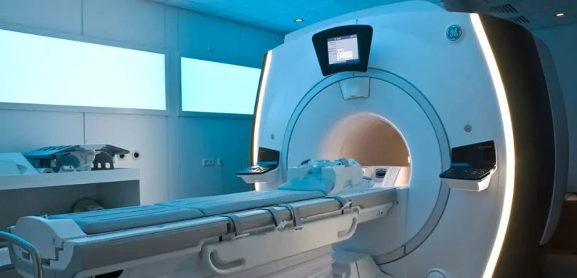
MRI in particular rewards learners who invest time in normal appearances. The same structure can look completely different depending on the sequence being used, the field strength, and the body region. Until normal becomes familiar, nothing else will feel solid.
This is why dedicated anatomy platforms are so valuable early on. Anatomymaster.com is one example of a site built specifically for this kind of structured anatomy learning. It organises anatomy by region and image type, which makes it much easier to build a systematic understanding rather than picking things up randomly. Spending time on a focused anatomy resource before jumping into pathology is one of the most efficient things a radiology learner can do.
Online Platforms Worth Bookmarking
Radiopaedia
If you only use one free radiology resource, make it Radiopaedia. It is peer-reviewed, constantly updated, and covers almost every area of imaging in real depth. You can look up a condition and find a clear explanation alongside real cases, which is exactly what you need when you come across something unfamiliar in study or clinical practice.
Radiopaedia works for beginners checking definitions and for more advanced learners exploring differentials and pattern recognition. It is not just a reference site — it is a case library, a teaching tool, and a reporting aid all in one. Many radiologists still use it daily even after years of experience.
IMAIOS
IMAIOS is particularly strong for cross-sectional anatomy learning. Its e-Anatomy tool lets you click through anatomical structures on real imaging studies, including MRI and CT, and see exactly where each structure sits in relation to everything around it. For learners who find static diagrams too abstract, this kind of interactive format is genuinely transformative.
If you are working through MRI anatomy of a particular region — the brain, the knee, the abdomen — IMAIOS gives you a way to explore it in real imaging rather than just reading about it. That direct visual engagement is how anatomy actually sticks.
MRIMaster.com
For MRI-focused learning, MRIMaster.com is a practical resource built around the real challenges that students and professionals face. It covers MRI protocols, planning, techniques, and anatomy in a focused and accessible way. Because it is built by someone who understands the day-to-day realities of MRI learning, it feels more targeted than a general textbook — which is often exactly what learners need.
Books That Are Still Worth Reading
In an age of online everything, books still have a role. A good textbook gives you structure and depth that scattered online browsing usually cannot replicate. The key is choosing the right ones rather than trying to read everything.
For CT
Fundamentals of Body CT remains one of the most recommended introductory texts for cross-sectional imaging. It connects images to anatomy clearly and gives learners a solid framework for understanding CT anatomy of the chest, abdomen, and pelvis. If you are serious about cross-sectional radiology, this is a foundational read.
For Chest X-Ray
Felson’s Principles of Chest Roentgenology is a genuine classic. Its programmed learning format makes it accessible even for complete beginners, and it has helped generations of learners build confidence with chest films. Another popular option is The Chest X-Ray: A Survival Guide, which is more concise, very image-heavy, and practical for daily use. Both are worth having.
For General Radiology
Brant and Helms is often the first recommendation for learners who want a broad overview of radiology before specialising. It covers multiple modalities and body systems, which makes it useful as an orientation text before you go deeper into one area.
For Pathology
Radiology makes much more sense when you understand the underlying disease process. Robbins Basic Pathology is not a radiology book, but pairing it with imaging study is a strategy that many strong learners use. When you understand why a tumour or infarct or inflammatory lesion behaves the way it does at a tissue level, the imaging appearances stop being arbitrary and start making biological sense.
YouTube Channels: A Detailed Guide
YouTube is one of the most underrated resources in radiology education. For a visual, image-based subject, being able to watch someone walk through a scan, explain a concept, or demonstrate a technique in real time is genuinely powerful. The key is knowing which channels are worth following.
MRI Physics and Techniques
Radiology Tutorials One of the most comprehensive free channels for MRI and CT education. Covers MRI physics, sequences, artefacts, and anatomy across a wide range of videos. It is methodical and well-organised, making it easy to work through topics systematically rather than randomly. Particularly strong for learners who want to understand why sequences look the way they do, not just what they look like.
MRI Physics with Nathan McDannold (Harvard) A series of university-level MRI physics lectures freely available on YouTube. Goes into deeper detail on signal generation, k-space, gradient echoes, and spin echoes. Ideal for radiographers sitting physics exams or anyone who wants a solid theoretical foundation to support their clinical work.
The Noted Anatomist Covers anatomy across multiple imaging modalities with clear, well-labelled walkthrough videos. Particularly useful for students combining anatomy study with cross-sectional imaging learning.
Radiopaedia YouTube Channel Radiopaedia has its own YouTube channel with case-based teaching videos, anatomy walkthroughs, and tutorials from experienced radiologists. A natural complement to the website — use the channel when you want a guided explanation rather than reading through articles.
Dr. Matt and Dr. Mike A popular anatomy and medical education channel that covers imaging anatomy alongside clinical medicine. Good for beginners who want an accessible introduction to how radiology connects to anatomy and clinical findings, without getting too technical too quickly.
CT Learning on YouTube
CTisus – Elliot Fishman One of the most respected names in CT education, Professor Elliot Fishman from Johns Hopkins has posted extensive lecture-style videos covering CT technique, anatomy, pathology, and contrast use. The content is aimed at radiologists and trainees but is accessible to serious learners at any level. Particularly strong for abdominal and oncological CT.
Frank Gaillard / Radiopaedia Lectures Several lecture series from Radiopaedia contributors cover CT interpretation systematically, with worked examples and pattern-recognition teaching. These videos are especially useful for building reporting confidence alongside the Radiopaedia case library.
Radiology Nation Covers practical CT and MRI interpretation with a clinical focus. The videos are designed for trainees and practitioners who want to improve their real-world image reading rather than just their theoretical knowledge. Useful for chest CT, abdominal CT, and neuroimaging.
X-Ray and General Radiology
Geeky Medics Excellent for medical students approaching radiology for the first time. Covers chest X-ray, abdominal X-ray, and basic imaging interpretation in a structured, exam-focused format. The explanations are clear and the pace is suitable for beginners.
Armando Hasudungan A widely loved medical illustration and teaching channel. While not exclusively radiology, the anatomy and pathology videos are consistently high quality and pair well with imaging study. The visual style makes complex concepts much easier to absorb.
MRI Sequences and Protocols
Questions and Answers in MRI (allaboutMRI) Based on the excellent website of the same name, this channel explores specific MRI sequences, signal characteristics, and clinical applications in short, focused videos. If you have ever wondered why a particular sequence is chosen for a clinical question, this channel gives real answers. It covers topics like STIR, FLAIR, DWI, gradient echo, and many others in plain language.
MRI Online A professional MRI education platform with a YouTube presence offering free content on sequences, protocols, and clinical applications. The paid platform goes deeper, but the free YouTube videos are genuinely useful for getting an overview of MRI techniques and understanding how different sequences contribute to a diagnosis.
MRI Safety Resources: Essential and Often Overlooked
MRI safety is one of the most important and most underemphasised areas in radiology education. Every person working in or near an MRI environment — radiographers, radiologists, nurses, porters, anaesthetists — needs a clear understanding of the risks and protocols involved. The consequences of MRI safety failures can be severe, which is why this area deserves its own dedicated attention.
Key Websites for MRI Safety
MRIsafety.com This is the most widely used free MRI safety reference in the world. It contains a searchable database of implants, devices, and materials tested for MRI compatibility, along with detailed safety information for each. Before scanning any patient with an implant or device, checking MRIsafety.com should be standard practice. It also includes educational articles, guidelines, and case reports covering a wide range of safety topics.
The Institute for Magnetic Resonance Safety, Education, and Research (IMRSER) IMRSER publishes peer-reviewed guidance on MRI safety, including recommendations for screening patients, managing implants, and safe working practices in the MRI environment. Their materials are referenced in clinical guidelines and are useful for anyone wanting to understand the evidence base behind safety protocols.
American College of Radiology (ACR) MRI Safety Guidelines The ACR publishes regularly updated MRI safety guidance documents covering patient screening, contrast agents, acoustic noise, pregnancy, and implant management. These are authoritative, comprehensive, and freely available on the ACR website. They are the standard reference for MRI safety practice in many countries.
British Institute of Radiology (BIR) and MHRA (UK) For UK-based learners and practitioners, the Medicines and Healthcare products Regulatory Agency (MHRA) publishes safety guidelines for MRI equipment and patient management. The BIR also provides educational resources and professional guidance relevant to MRI practice in the UK.
MRI Safety on YouTube
MRI Safety Education – Frank Shellock Frank Shellock is one of the most respected names in MRI safety research and is closely associated with MRIsafety.com. Lecture recordings and presentations from Shellock are available online and provide an authoritative overview of safety issues, implant testing, and practical screening protocols.
SMRT (Society for MR Radiographers and Technologists) SMRT has educational video content covering MRI safety, patient care, and professional practice for MRI technologists. Their materials are especially useful for radiographers working towards professional development or preparing for safety-focused assessments.
Radiology Cafe and Similar Trainee Channels Various radiology trainee and radiographer channels on YouTube cover MRI safety topics alongside clinical teaching. While these are less formal than institutional resources, they often present safety concepts in a very accessible way that works well for learners encountering them for the first time.
Key MRI Safety Topics Every Learner Should Cover
Whatever resources you use, make sure you understand these core areas:
The four MRI safety zones (Zones I–IV) and what each means for access and patient management. This is the foundation of safe MRI practice.
Ferromagnetic projectile risk — understanding why loose metallic objects near an MRI room are dangerous, and how to prevent accidents.
Implant and device screening — how to assess whether a patient’s implant is MRI conditional, MRI safe, or MRI unsafe, and what additional precautions may be needed.
Contrast agents and nephrogenic systemic fibrosis (NSF) — the risks associated with gadolinium-based contrast agents in patients with renal impairment, and current guidelines on contrast use.
Acoustic noise — understanding the hearing risk associated with MRI scanning and appropriate use of hearing protection.
Pregnancy and MRI — current guidance on scanning pregnant patients, including first trimester considerations and gadolinium use.
Thermal effects and SAR (Specific Absorption Rate) — understanding how radiofrequency energy is deposited in tissue and how SAR limits protect patients.
How to Put It All Together
The most effective radiology learners use a layered approach rather than relying on one source. Here is a simple structure that works well:
Books for structure — one general text, one modality or region-specific text, and MRI in Practice for physics.
Anatomy sites for orientation — Anatomymaster.com and IMAIOS for working through normal anatomy before studying pathology.
Case libraries for pattern recognition — Radiopaedia is the obvious choice. Review cases regularly, not just when looking something up.
YouTube for reinforcement — after reading a topic, watch one of the channels listed above to make the concept concrete. Radiology Tutorials for general physics, CTisus for CT, Questions and Answers in MRI for sequences, and MRIsafety resources for safety.
MRIMaster.com for MRI-specific practice — protocols, planning, and technique details that general texts often do not cover well.
Safety resources as a constant companion — MRIsafety.com and the ACR guidelines should be consulted regularly, not just read once.
Free vs Paid: Getting the Balance Right
One of the most reassuring things about radiology learning today is how much is available for free. Radiopaedia is free. YouTube is free. MRIMaster.com offer substantial free content. MRIsafety.com is free. The ACR guidelines are free. You do not need to spend a lot to build a strong foundation.
Paid resources — premium anatomy atlases, structured online courses, digital textbooks — can be worth the investment once you know what you need. But for most learners, especially early on, the free resources available today are genuinely excellent.
Summary
There is no single perfect resource that will teach you everything. Radiology learning works best when you combine a few strong, complementary tools and return to them consistently. Anatomy first, then pathology, then reporting patterns. YouTube channels bring physics and technique to life. MRI safety resources are not optional — they are part of the job.
The combination of MRIMaster.com, Anatomymaster.com, Radiopaedia, IMAIOS, a solid physics textbook, and the YouTube channels listed above will take most learners further than any single expensive course. Build the habit of consistent, layered learning and the subject will reward you for it.